삼성서울병원 2025년 및 2026년 임상강사 여러분께
2026년 제가 계획하고 있는 여러 프로그램을 안내합니다.
1. 1월 첫 주에 ESD 2026 (Endoscopy Samsung Days 2026)를 개최합니다. 수요일과 금요일은 원내 세미나이고 토요일 오전은 신논현역 인근에서 초청연자와 내부 연자를 모시고 세미나를 하고 점심식사를 한 후 해산할 것입니다.
2. [임상시뮬레이션센터와 함께하는 Saturday morning on-line endoscopy seminar 2026]를 3월 7일 토요일과 9월 5일 토요일에 진행합니다.
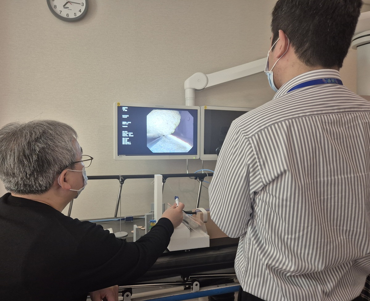
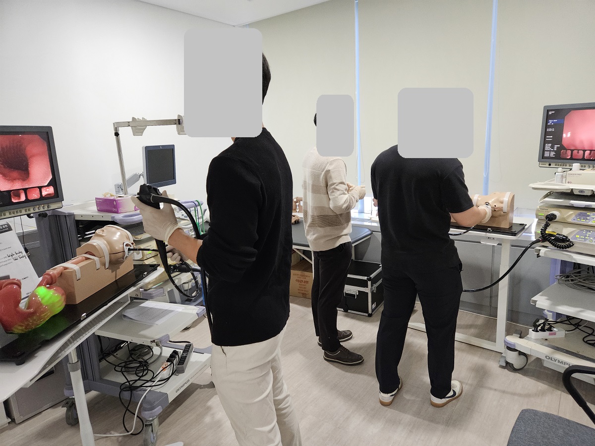
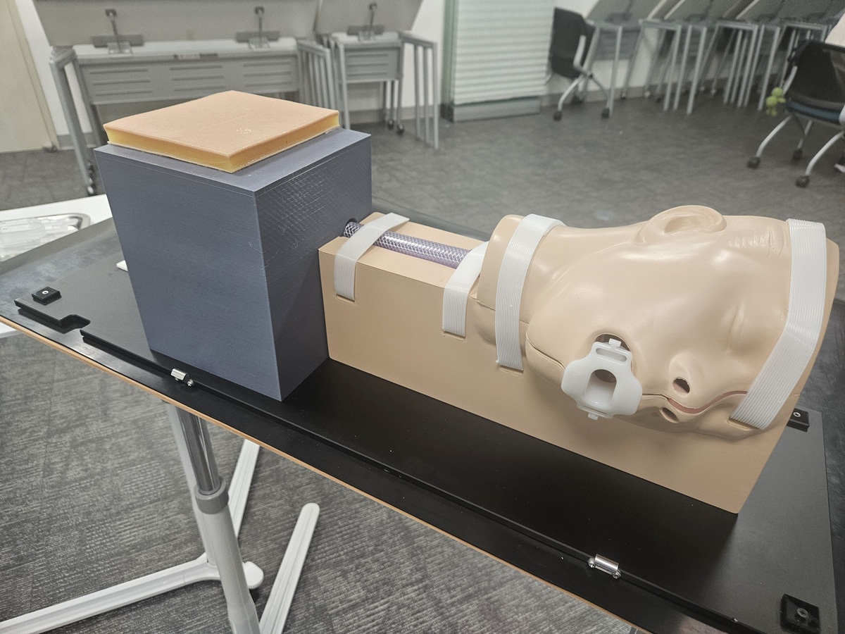
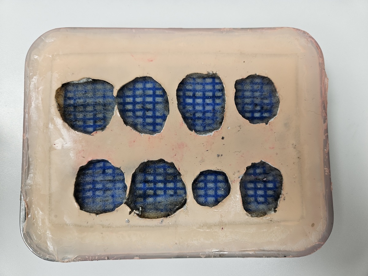
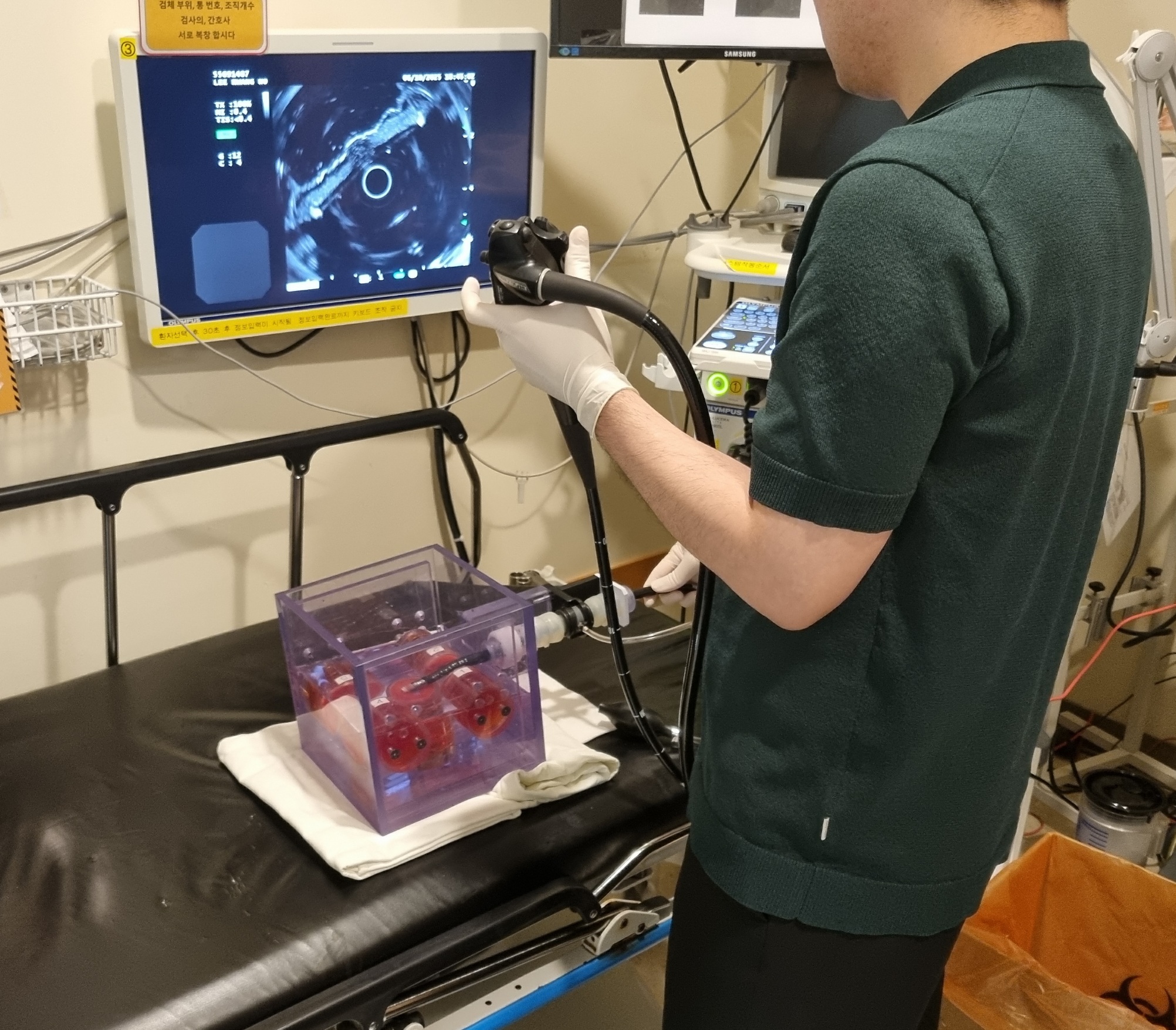
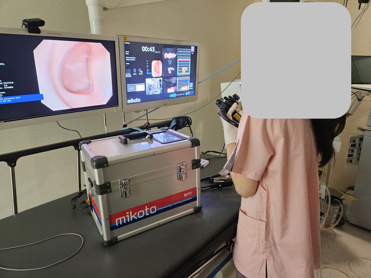
3. 임상시뮬레이션센터와 함께하는 2026년도 Hands-on training 계획(案)도 잡았습니다. 물론 원내의 별도 hands-on도 별도로 많이 준비될 것입니다.
4. The MOST라는 잡지에 "소화기질환 진단 및 치료에 관한 내시경 강좌"를 매월 기고하려고 합니다. 리뷰와 증례와 Q and A와 동영상 lecture로 구성된 흥미로운 자료가 될 것입니다.
5. 2026년 월간 sponsored 세미나도 마련하였습니다. 제약사의 세미나도 내용이 좋을 수 있음을 보여줄 것입니다. 제가 좌장이나 연자를 하고 몇 몇 분을 초대할 것입니다. 함께 가시고 싶은 분은 미리 알려주세요. 소주 한 잔 대접하겠습니다.
6. 이준행 교수와 함께하는 학회 참석 일정도 미리 알려드립니다. 밥 사겠습니다.
7. 2026년 1년차 fellow를 위한 DEX quiz 풀이는 이미 시작하였습니다.
이준행
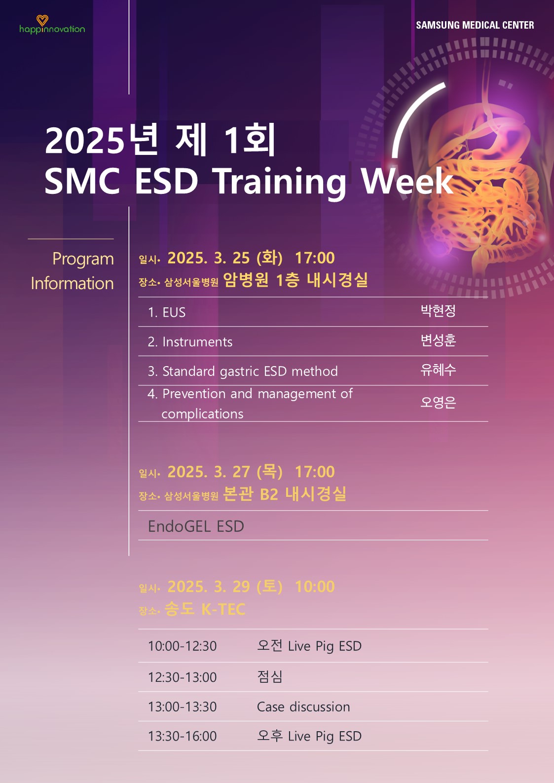
| ESD 2026 (Endoscopy Samsung Days 2026) |
| 1월 7일 수요일 7:30-8:00 | 이준행 특강 (TBA) | SMC 암병원 내시경 회의실 | 식사 없음 |
| 1월 9일 금요일 8:00-9:00 | 병리집담회 | SMC 암병원 내시경 회의실 | 식사 없음 |
| 1월 10일 토요일 9:00-11:00 | Special lecture (이화여자대학교 태정현 교수)와 증례 토의 (2) | 신논현역 인근 | 점심 식사 후 해산 |
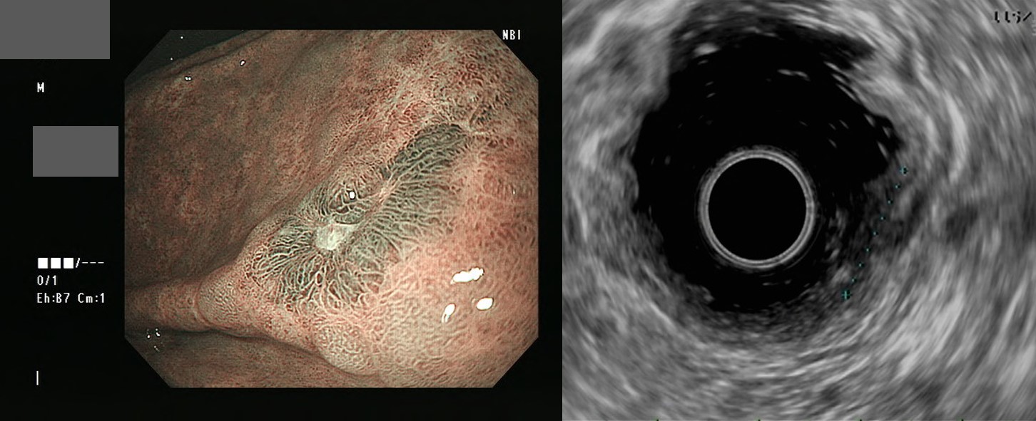
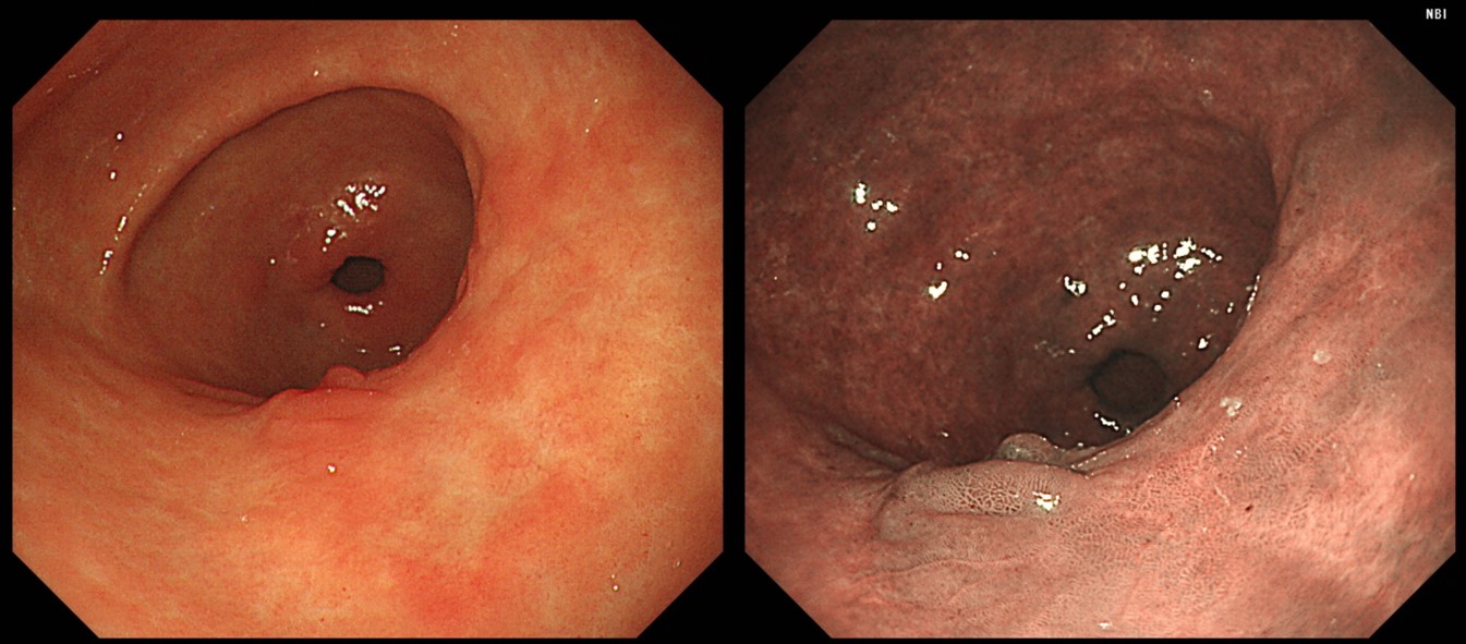
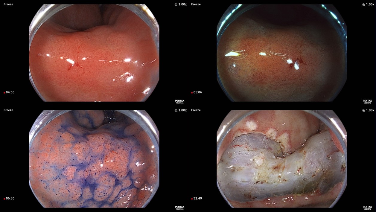
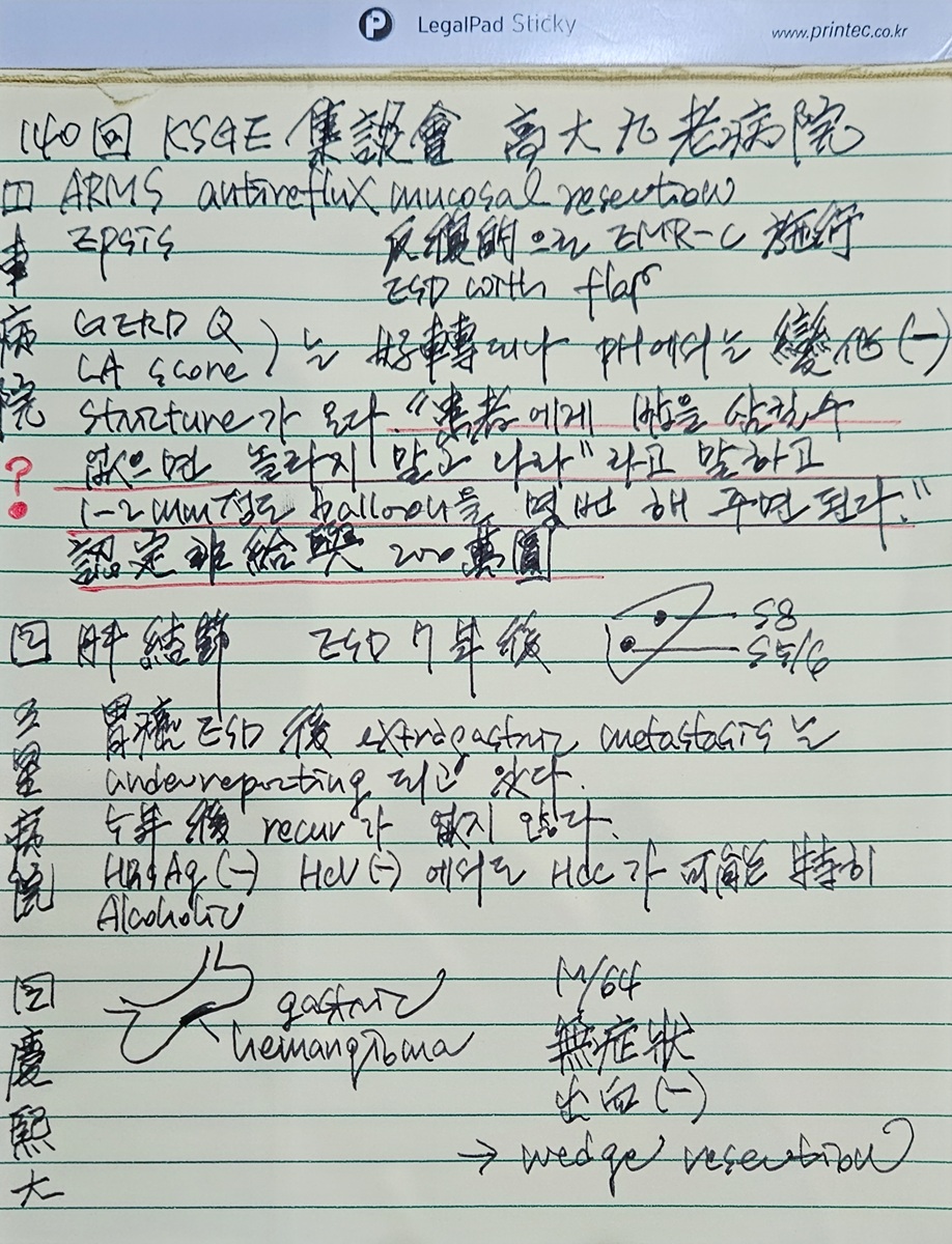
[증례 토의 목록]- 오영은 - Gastric cancer in juvenile polyposis syndrome (지도교수: 이준행)
- 이상훈 - ESD for Barrett adenocarcinoma (지도교수: 이준행) 1-2025-03
|
[임상시뮬레이션센터와 함께하는 Saturday morning on-line endoscopy seminar 2026]
대상: 소화기내과 임상강사, 내과 전공의, 그리고 원내외 원하시는 모든 분
Concept: 제목: 전공의, 임상강사, 개업의를 위한 내시경 진단과 치료의 실제. 교육인재개발실과 함께 프로그램을 개발하여 토요일 오전 zoom으로 진행하는 공개 강의 프로그램. 이론보다는 실무를 강조. 되도록 많은 증례를 토의. 하루에 4시간 (내시경 공통 1시간, 상부 2시간, 하부 1시간). 강사의 강의 후 좌장과 함께 많은 증례를 토의하고 질문에 답하는 형식. 원내 무료, 원외 유료. 소화기내과 Winter school program과 겹치지 않도록 적절한 주제 선정
[2026년 3월 7일 (안) 9:00-13:00] 전공의, 임상강사, 개업의를 위한 내시경 진단과 치료의 실제. 좌장: 이준행
- 내시경 진정과 조직검사 (민병훈)
- 위궤양과 위암의 감별 진단 (이혁)
- Helicobacter 감염의 진단과 치료 (김태세)
- 대장 용종과 조기 대장암 (김은란)
[2026년 9월 5일 (안) 9:00-13:00] 전공의, 임상강사, 개업의를 위한 내시경 진단과 치료의 실제. 좌장: 이준행
- 조기위암 내시경 치료. 어떻게 시작할 것인가? (김태세)
- 식도암의 진단과 치료. 내시경 치료를 중심으로 (민양원)
- 상부위장관 출혈의 내시경 치료 (오영은)
- 소장내시경 (홍성노)
[2026년도 Hands-on training 계획(案)]
- 2026년 3월 28일 토 오전 Colonoscopy-BOXIM 김은란 (원내 한)
- 2026년 3월 28일 토 오후 Colonoscopy-BOXIM 김지은 (원내 한)
- 2026년 4월 14일 화 18:30 DEX basic 이준행/김태세
- 2026년 4월 28일 화 18:30 DEX basic 이준행/김태세
- 2026년 4월 25일 토 오전 G-BOXIM 김태세
- 2026년 4월 25일 토 오후 G-BOXIM 오영은
- 2026년 5월 23일 토 오전 G-BOXIM 민병훈
- 2026년 5월 23일 토 오후 G-BOXIM 변성훈
- 2026년 6월 27일 토 오전 Colonoscopy-BOXIM 김민지
- 2026년 6월 27일 토 오후 Colonoscopy-BOXIM 김경완
- 2026년 9월 19일 토 오전 Colonoscopy-BOXIM 변성훈
- 2026년 9월 19일 토 오후 Colonoscopy-BOXIM 박현정
- 2026년 9월 22일 화 ESD 김태세 (원내 한)
- 2026년 10월 6일 화 PEG 김태세 (원내 한)
- 2026년 10월 13일 화 18:30 DEX basic 이준행/김태세
- 2026년 10월 27일 화 18:30 DEX basic 이준행/김태세
- 2026년 10월 24일 토 오전 G-BOXIM 이혁
- 2026년 10월 24일 토 오후 G-BOXIM 김경완
- 2026년 11월 3일 화 POEM 민양원 (원내 한)
- 2026년 11월 28일 토 오전 G-BOXIM 김태세
- 2026년 11월 28일 토 오후 G-BOXIM 박현정
[The MOST] 소화기질환 진단 및 치료에 관한 내시경 강좌 연중 기획
| 소화기질환 진단 및 치료에 관한 내시경 강좌 |
1. A4 용지 3-4페이지 종설 (여러분) - 전달 10일까지 이준행에게 원고를 전달해 주시기 바랍니다. 2. 내시경 증례 (이준행) 3. FAQs (다함께) - 가장 궁금한 질문 5개를 보내주세요. 이준행이 답을 준비하겠습니다. 4. On-line lecture (이준행) YouTube 강의를 녹화하여 Medical Observer 사와 이준행 YouTube에 동시에 올리기. 원고에는 QR 코드를 공개할 예정입니다. |
1월: 실전 위암과 위궤양의 감별진단 (박현정) 2월: 실전 조기위암의 내시경 진단과 치료 (김경완) 3월: 실전 조기위암 내시경 치료 후 관리 방안 - 국소재발, 이소성재발, 헬리코박터 제균치료 (김태세) 4월: 실전 위선종의 내시경 진단과 치료 (변성훈) 5월: 실전 출혈성 위궤양의 진단과 치료 (박현정) 6월: 실전 위식도역류질환의 내시경 소견과 약물치료 (오영은) 7월: 실전 단일 미란, 불응성 궤양의 진단과 치료 (김경완) 8월: 실전 식도 이물의 진단과 치료 (오영은) 9월: 실전 헬리코박터 감염의 진단과 치료 (변성훈) 10월: 실전 위 MALT 림프종의 진단과 치료 (2026년 2년차) 11월: 실전 위축성 위염, 화생성 위염, 자가면역위염 (2026년 2년차) 12월: 실전 다양한 임상상황에서의 상부위장관 출혈 (2026년 2년차) |
[2026년 월간 sponsored 세미나]
- 2월 9일 (월) 알쏭달쏭
- 3월 16일 (월) EndoTODAY
- 4월 13일 (월) GERD
- 5월 18일 (월) 알쏭달쏭
- 6월 15일 (월) EndoTODAY
- 8월 18일 (화) 알쏭달쏭
- 9월 14일 (월) EndoTODAY
- 10월 12일 (월) GERD
- 11월 2일 (월) 알쏭달쏭
- 11월 9일 (월) EndoTODAY
[이준행 교수와 함께하는 학회 참석]
- 2026년 3월 28일 (토) KGCA 위암학회 연수강과 (대구) - 저는 금요일에 내려갔다가 토요일 저녁에 올라올 예정입니다.
- 2026년 5월 7일 (목) - 9일 (토) 일본내시경학회 (요코하마) - 저는 목요일 오후 6시 경 비행기로 출국하여 토요일 저녁 비행기로 귀국할 예정입니다.
- 2026년 7월 18일 (토) - 19일 (일) 요코하마 라이브 & 일본 EMR/ESD 연구회 (요코하마) - 저는 금요일 밤 출국하여 일요일 밤 귀국할 예정입니다.


 2025-1-11


