
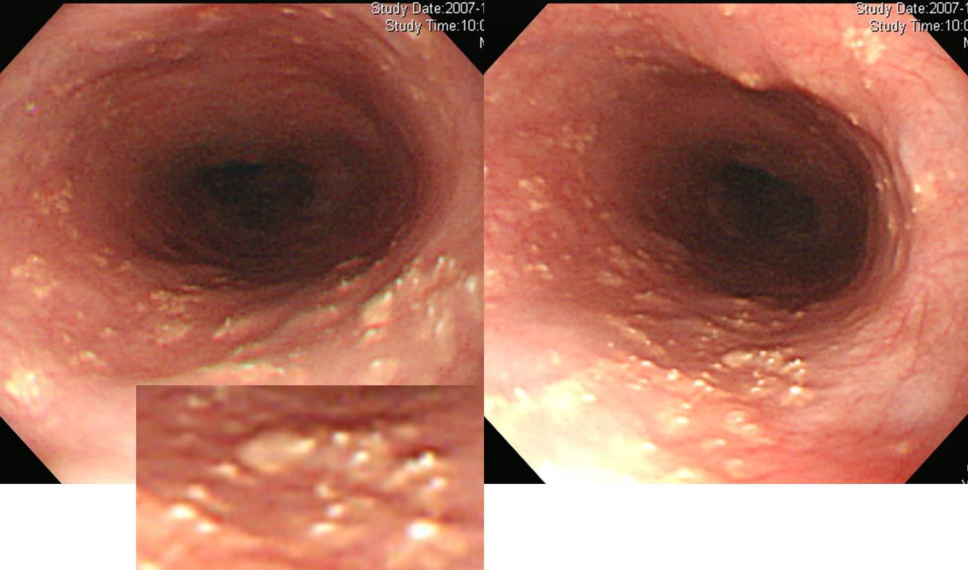
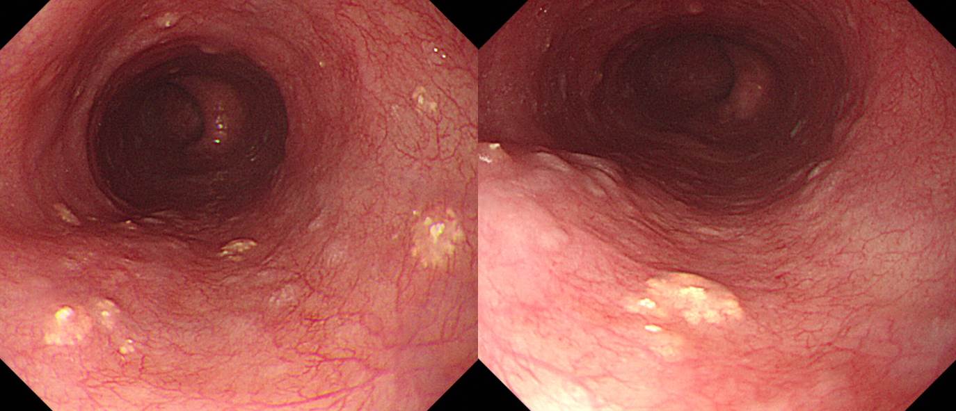
Ectopic sebaceous gland는 다수의 작은 연노란색 plaque이면서 그 중앙에 매우 작은 흰색 papule이 보이는 병소로 관찰됩니다. Congenital인지 acquired인지 불분명하여 sebaceous gland metaplasia라고 부르기도 합니다. 참고문헌을 옮깁니다.
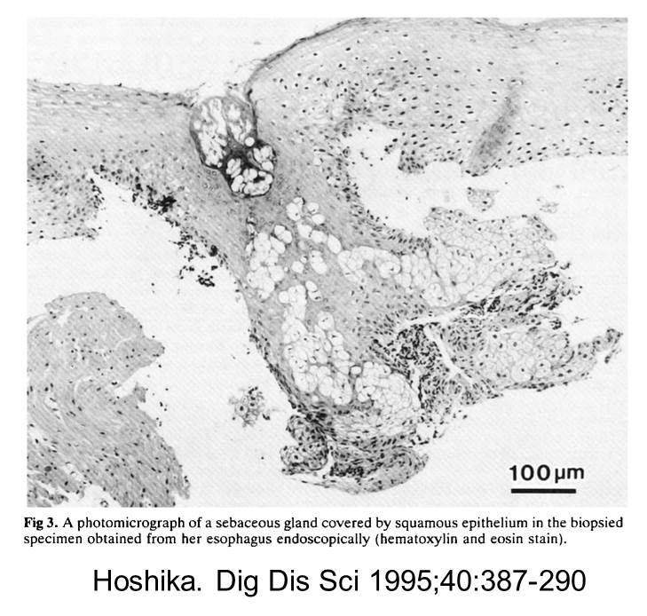
Sebaceous glands generally arise in close association with hair follicles to form the pilosebaceous apparatus, but they are also found independently. Ectopic sebaceous glands have been reported arising at a variety of sites, including oral cavatiy, genitals, eye and orbits, nipples, palms and soles, parotid glands, and esophagus. Ectopic sebaceous glands in the esophagus are recognized as one or more small maculas in the esophageal mucosa and resemble gastric xanthomas. (Hoshika. Dig Dis Sci 1995;40:387-290)
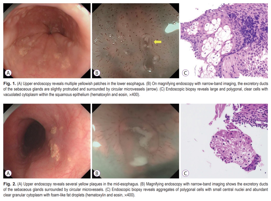
식도피지선은 상피하의 점막고유층에 존재하고 있고 점막고유층의 선방(腺房)과 식도내강으로의 외분비도관으로 이루어져있다. 따라서 육안소견은 작은 황백색조의 편평융기라든지 과립상의 요철을 수반하는 꽃잎모양 융기 등으로 관찰되는 선방부분과, 중심부나 정상에 백색의 작은 돌기가 보이는 도관부분으로 구성되어 있다. 위 xanthoma와 유사한 황색조의 융기성 병변으로 관찰되는데, 도관부분인 중심의 작은 원형의 황색점이라든가 작은 융기가 보이면 피지선(sebaceous gland)으로 진단할 수 있다. (Monna. 위와 장. 2008;43:263-273)
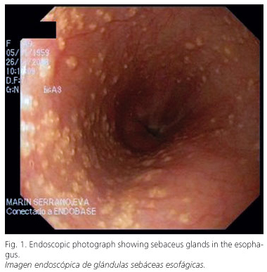
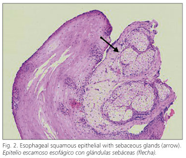
The discovery of sebaceous glands in ectoderm-derived tissues such as the oral cavity, parotid glands, and external genitalia is a frequent finding. On the contrary, the presence of sebaceous glands in the esophagus, which is an organ of endodermal origin, is rare: there are fewer than thirty cases reported, most of them affecting the esophagus in a patchy pattern. A 50-year-old woman suffering from heartburn and acid regurgitation was referred to our department for endoscopic examination. Endoscopy demonstrated more than one hundred tiny, rounded, elevated, white-yellowish lesions distributed throughout the entire esophagus, which were identified during the histopathological examination as sebaceous glands. As of now, the histogenesis of ectopic sebaceous glands in the esophagus is unknown; whilst it could be a congenital abnormality, a majority of authors defined it like an acquired metaplastic process. From a practical standpoint, it is an incidental finding that on occasion, as in our case, has been associated with gastroesophageal reflux disease, not requiring treatment or endoscopic follow-up and, and needing a differential diagnosis from Candida infection and glycogen acanthosis.
2018년 Clin Endosc에 ectopic sebasecous gland의 확대내시경 소견이 발표되었습니다. Duct가 보인다고 합니다.

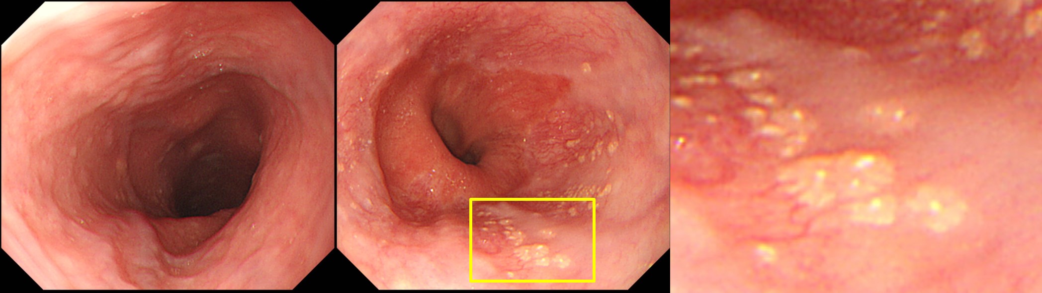
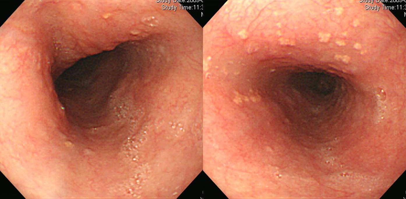
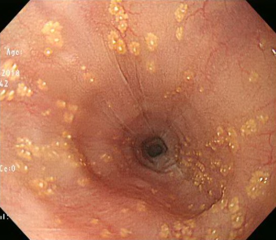
Reflux esophagits LA-B로 의뢰되었으나 자세히 관찰한 바 하부식도에 국한된 이소성 피지샘이었습니다. 뚜렷한 mucosal break는 없었습니다.
이 정도 전형적이면 조직검사도 필요하지 않습니다.

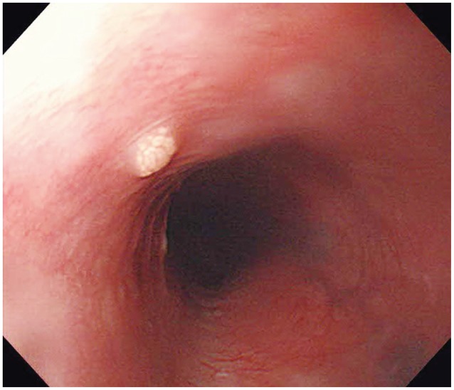
Esophageal xanthoma와 구분이 필요합니다. 식도에 xanthoma가 없는 것은 아니지만 너무 드물어 case report 수준입니다. 아래는 한 case report (Clin Endosc 2014)의 esophageal xanthoma 사진입니다.
esophageal xanthoma
[2018-2-14. 애독자 질문]
전임의 시절부터 EndoTODAY 구독을 시작해서 현재 2차병원 봉직의로 일하고 있습니다. 항상 가르침 감사합니다. 식도 병변에 대한 질문이 있어 메일 드립니다.
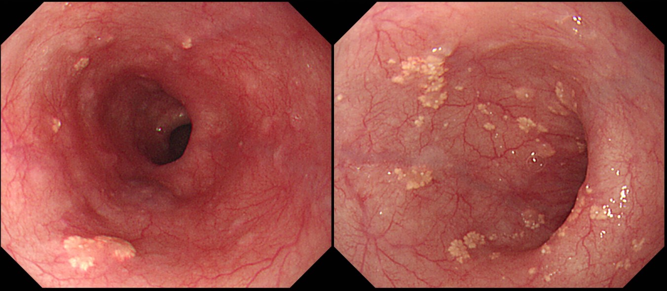
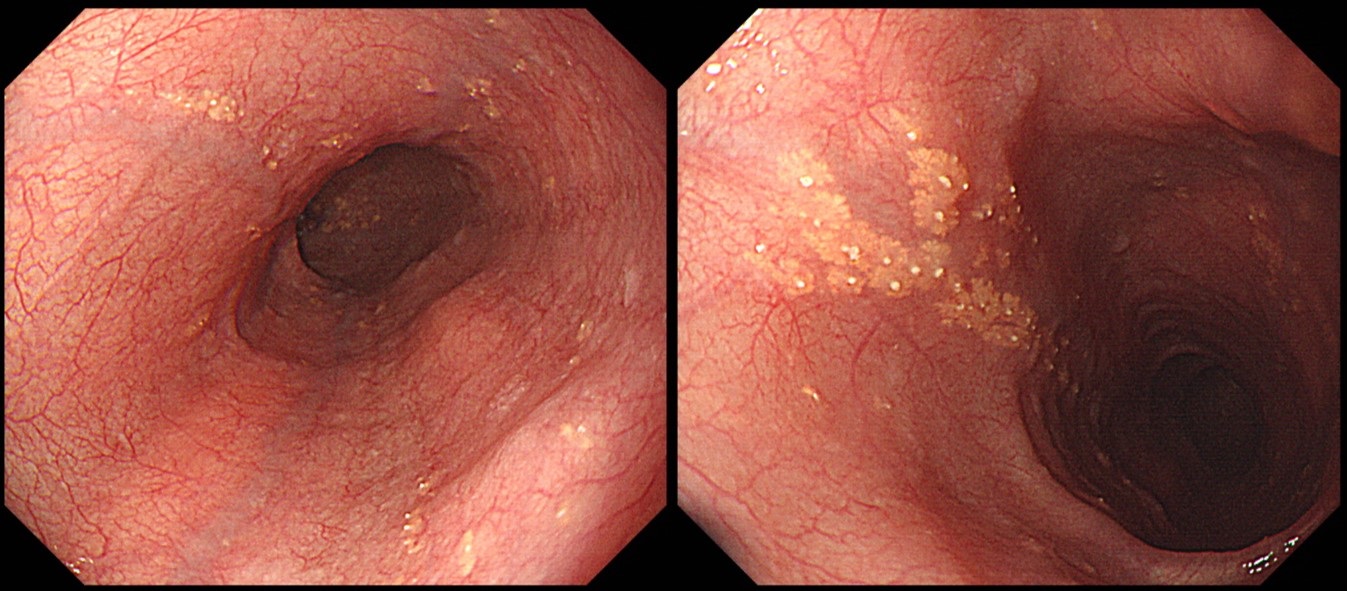
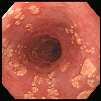
검진 내시경상 중부 식도부터 하부식도까지 다수의 작은 황색판이 관찰된 환자가 있었습니다. 세척에도 씻기지는 않는 병변이었고, 해당 부위에서 생검을 시행을 하였습니다. 환자 분께 증상은 못여쭤 봤으나 기록상에 투약력 등의 과거력은 없는 분이었습니다. Candidiasis는 아닐 것으로 생각되는데... 아틀라스에도 비슷한 병변은 보이지 않고...
혼자 고민을 해도 주변 선생님들하고 이야기를 해보아도 결론을 내지 못하여 메일 드립니다.
[2018-2-15. 이준행 답변]
[2018-11-24. 애독자 질문]
식도 cadidiasis grade I과 ectopic sebaceous gland의 구분이 어렵습니다. 둘 다 사진만 찍고 기술하지 않는 경우가 대부분이지만... 또는 어차피 치료하지 않을 거라 환자가 사진을 보고 물어보는 경우는 ectopic sebaceous gland 쪽으로 대답하기도 합니다.
[2018-11-26. 이준행 답변]
경증 candidiasis가 ectopic sebaceous gland보다 훨씬 흔합니다. 애매하더라도 자신이 판단한 바대로 설명드리는 것이 좋겠습니다. 무증상 식도 칸디다증은 있다 없다 하는 상황이고, ectopic sebaceous gland는 쭉 계속해서 관찰되는 것이니 전혀 다른 것입니다.
Ectopic sebaceous gland는 다수의 작은 연노란색 plaque이면서 그 중앙에 매우 작은 흰색 papule이 보이는 병소로 관찰됩니다. 가까이 접근해서 관찰하면 칸디다와는 제법 다릅니다.
댓글 없음:
댓글 쓰기